Photoacoustic tomography – Variational Methods for Improving Spatial Resolution
Researchers: Simon Arridge, Martin Benning, Sarah Bohndiek, Joanna Brunker, Tamara Grossmann, James Joseph, Erlend Riis, Carola-Bibiane Schönlieb, Michal Tomaszewski,
Photoacoustic tomography (PAT) is a novel biomedical imaging technique for imaging optical absorption in biological tissue. Obtaining spatiotemporal distributions of optical absorption in tissue is of significant interest in physiology and medicine, for example for understanding the underlying processes of tumour growth and how the tumours respond to different drugs. However, purely optical imaging techniques are limited by the high scattering of photons in tissue, which restricts the imaging depth to about 1mm. On the other hand, ultrasound imaging can image tissue at several centimeters depth, because ultrasound waves propagate linearly through soft tissue, but they lack the high contrast and physiological specificity of optical imaging. PAT combines the advantageous properties of each of these modalities by exploiting a phenomenon known as the photoacoustic effect, thus overcoming the imaging depth limit of optical methods (Beard, 2011). Known for its ability to render high resolution images of tissue at several centimetres depth, PAT has grown in the last decade to become a highly active area of research, that holds great promise for clinical and pre-clinical studies of physiological processes.
PAT imaging deals with large data sizes: In order to achieve a high (sub-millimetre) spatial resolution with PAT, one typically needs to take ultrasound measurements at a rate of tens of millions samples per second (Dima et al, 2104), and from all angles of the surface of the imaging object. To make the reconstruction problem computationally feasible and efficient, it is common to use ultrasound sensors whose apertures are focused to an imaging plane, so that one can reconstruct in 2D slices instead of in 3D. Such a transducer system, for cross-sectional imaging of mice, is employed at Dr. Sarah Bohndiek’s Vision Laboratory at the CRUK (Rosenthal et al, 2010). However, in spite of the use of focused ultrasound sensors, acoustic signals arriving from outside of the imaging plane impact the measurements and the subsequent reconstructions, leading to a significant image blurring perpendicularly to the imaging plane. While a 3D reconstruction model could accurately correct for these artefacts, it is computationally infeasible. It is therefore of great interest to develop efficient techniques to correct for this out-of-plane blurring.
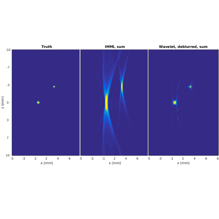
We are studying how to overcome these artefacts, through the combination of regularised reconstructions in a variational framework, and computationally cheap post-processing deconvolution steps (Buehler et al, 2014). Below, you can see a result from this work.
