Multi-modal image analysis in cancer research
Researchers: Kevin Brindle, Veronica Corona, Stefanie Reichelt, Carola-Bibiane Schönlieb
Multi-modal imaging has become increasingly important in medicine and biology, representing
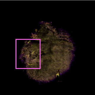
an essential tool to link anatomical and functional information. New developments in imaging technology, especially in light microscopy, make possible imaging of a whole sample at high spatial resolution. In this work, we want to correlate MRI and light microscopy images, where MRI gives geometrical information on shape and macroscopic structures of organs and tumours, while microscopy reveals inner structures such as blood vessel organisation or protein expression (Breckwoldt, 2016).
In this project, we are interested in developing new analysis methods for the correlation of MRI and microscopy data. Linking these two techniques allows us to understand the contrast mechanism in MRI, where microscopy acts as a ground truth of what it is truly hidden inside each voxel. This is especially useful in the assessment of tumour response to treatments at an early stage, allowing doctors to rapidly change strategy in the case of negative response.